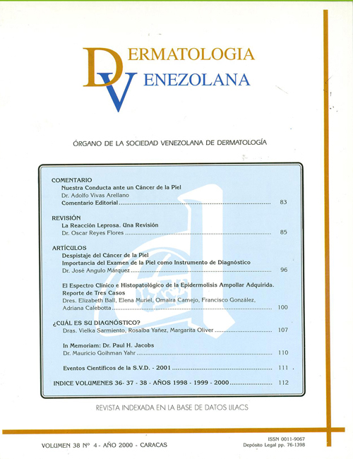
EL ESPECTRO CLÍNICO E HISTOPATOLÓGICO DE LA EPIDERMOLISIS AMPOLLAR ADQUIRIDA. REPORTE DE TRES CASOS
Palabras clave:
Ampollas subepidérmicas, Epidermolisis ampollar adquirida, Separación cutánea por NaCI, Epidermolysis Bullosa Acquisita, Salt-Split Skin Methods, Subepidermal BullaeResumen
La epidermolisis ampollar adquirida (EAA) es una enfermedad ampollar subepidérmica, crónica, poco frecuente, caracterizada por la presencia de autoanticuerpos circulantes y/o tisulares tipo IgG dirigidos contra el colágeno tipo VII de las fibrillas de anclaje de la unión dermo-epidérmica. Su espectro clínico e histopatológico es variable y se han definido tres presentaciones: la forma clásica similar a una porfiria cutánea tarda o a una epidermolisis ampollar distrófica, la forma tipo penfigoide ampollar y la forma tipo penfigoide cicatricial. El diagnóstico definitivo se establece mediante la inmunofluorescencia directa con piel perilesional separada con NaCI 1 molar o mediante la inmunomicroscopía electrónica. En nuestro medio, es una enfermedad poco frecuente o no es detectada ya que plantea dificultades diagnósticas con otras enfermedades ampollares. En este estudio presentamos tres casos catalogados como EAA recolectados de la consulta de enfermedades ampollares de nuestro centro, que ilustran la diversidad clínica e histológica de esta entidad.Clinical and Pathologic Spectrum of Epidermolysis Bullosa Acquisita. Report of three cases.
ABSTRACT
Epidermolysís bullosa acquisita is a chronic, infrequent, subepidermal blistering disease characterized by the presence of circulating and/or tissue-bound IgG autoantibodies directed against type VII collagen within anchoring fibrils structures that are located at the dermo-epidermal junction. The clinical and histologic spectrum of the disease is variable and there are at least three clinical variants: the classic presentation reminiscent of porphyria cutanea tarda or hereditary dystrophic epidermolysis bullosa, the bullous pemphigoid-like form and the cicatricial pemphigoid-like presentation.The diagnosis is established by direct salt-split skin inmunofluorescence of perilesional skin or by immunoelectron microscopy. In our country it is an infrequent or undetected disease, mainly because of the difficulties in the differential diagnosis with other subepidermal blistering diseases. In this study we report three cases of EAA from the clinic of blistering diseases of our hospital, which reflectthe clinical and histologic diversity of this entity.
Descargas
Número
Sección
Artículos
Licencia
Publicado por la Sociedad Venezolana de Dermatología Médica, Quirúrgica y Estética


